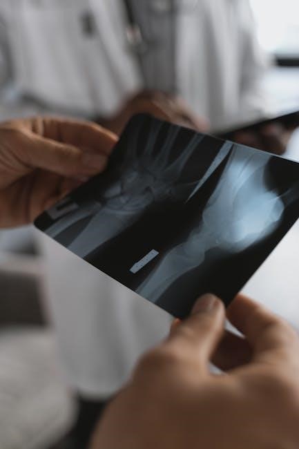
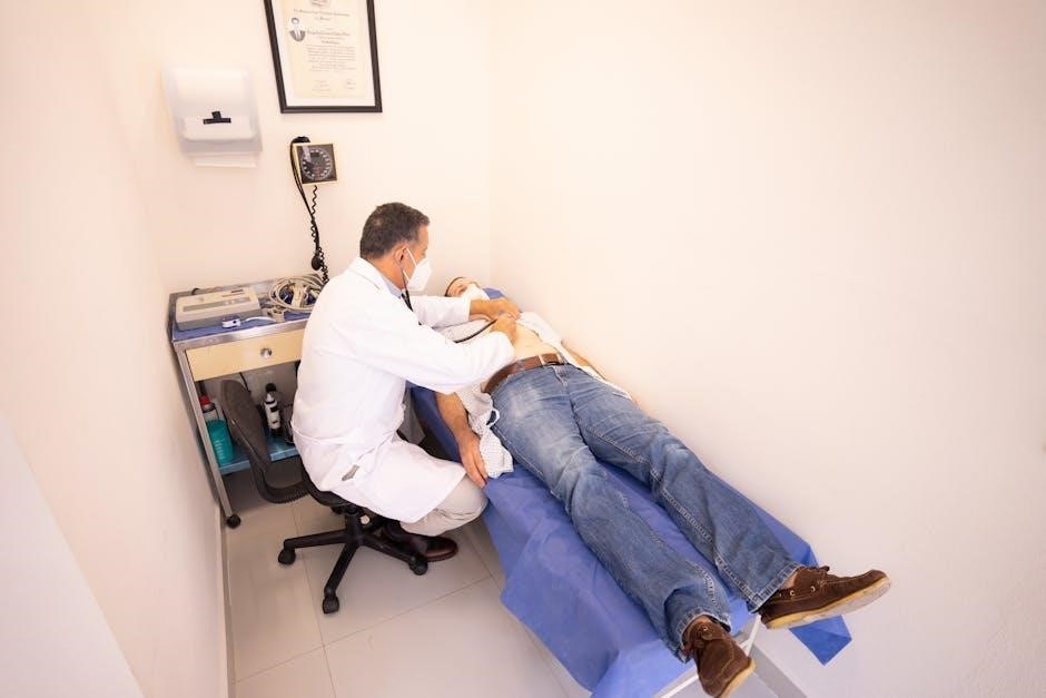
The physical examination of the musculoskeletal system is essential for assessing function, identifying abnormalities, and guiding treatment. It involves inspection, palpation, range of motion, and muscle strength testing to evaluate joints, muscles, and ligaments. This systematic approach helps clinicians diagnose conditions affecting mobility and pain, ensuring effective patient care and rehabilitation strategies.
Importance of Musculoskeletal Examination in Clinical Practice
The musculoskeletal examination is a cornerstone of clinical practice, enabling early detection of abnormalities, guiding treatment plans, and improving patient outcomes. It helps identify structural and functional deficits, distinguishing between acute and chronic conditions. Accurate assessment informs rehabilitation strategies, reduces recovery time, and enhances quality of life. Regular musculoskeletal evaluations are vital for preventing complications and ensuring optimal management of orthopedic and rheumatological disorders. This systematic approach is indispensable for clinicians in delivering targeted and effective care.
Overview of the Musculoskeletal System
The musculoskeletal system comprises bones, joints, muscles, ligaments, and tendons, enabling movement, stability, and support. It is divided into the axial (skull, spine, ribs) and appendicular (upper and lower limbs) components. This system facilitates locomotion, maintains posture, and protects internal organs. Understanding its structure and function is crucial for accurate physical examinations and diagnosing conditions such as arthritis, fractures, or soft tissue injuries. A thorough knowledge of this system aids in effective clinical assessment and treatment planning for musculoskeletal disorders.
Preparation for the Examination
Proper preparation is essential for an effective musculoskeletal examination. Ensure the patient is in a comfortable, private setting with adequate exposure of the relevant body areas. A quiet, well-lit room facilitates accurate assessment. Tools such as a reflex hammer and goniometer may be needed for specific tests. The examiner should be familiar with the sequence of examination to ensure efficiency. Mastery of the technique allows the exam to be completed in under 5 minutes, making it easy to integrate into the broader physical assessment. Proper preparation enhances diagnostic accuracy and patient trust.
The GALS (Gait, Arms, Legs, Spine) Screening
The GALS screening is a quick, reasonably sensitive method to detect common musculoskeletal abnormalities, focusing on gait, arms, legs, and spine assessment. It is a key component of a broader medical evaluation.
Gait Assessment
Gait assessment is a critical component of the musculoskeletal examination, evaluating the patient’s ability to walk and maintain balance. Observing the patient’s stance, stride, and movement patterns helps identify abnormalities such as limping, asymmetry, or difficulty initiating gait. Key elements include examining the phases of gait (stance and swing) and noting any deviations or compensatory mechanisms. Pain or limited mobility during walking may indicate underlying pathologies, such as joint degeneration or muscle weakness. This assessment provides valuable insights into functional mobility and guides further evaluation or therapeutic interventions.
Arm and Hand Examination
The arm and hand examination evaluates the upper limb’s musculoskeletal integrity, focusing on joints, muscles, and ligaments. Key steps include inspection for swelling or deformity, palpation for tenderness or warmth, and assessment of range of motion in the shoulder, elbow, wrist, and fingers. Muscle strength testing and special maneuvers, such as the Phalen’s test for carpal tunnel syndrome, are also performed. This systematic approach helps identify conditions like arthritis, tendonitis, or ligament sprains, guiding appropriate diagnostic and therapeutic interventions.
Leg and Foot Examination
The leg and foot examination involves a thorough assessment of the lower limb, focusing on joints, muscles, and ligaments. Key components include inspection for swelling or deformities, palpation for tenderness or warmth, and evaluation of range of motion in the hips, knees, ankles, and toes. Muscle strength testing and special maneuvers, such as the McMurray’s test for meniscal injuries, are also performed. This systematic approach helps identify conditions like arthritis, ligament sprains, or plantar fasciitis, ensuring accurate diagnosis and targeted treatment plans.
Spine and Posture Evaluation
Spine and posture evaluation is crucial for identifying musculoskeletal abnormalities. The assessment begins with inspection of posture, noting any deviations like kyphosis or scoliosis. The examiner checks for deformities, swelling, or asymmetry and evaluates spinal flexibility during forward bending, extension, and lateral flexion. Palpation is used to detect tenderness or muscle spasms. Postural alignment and gait are observed to identify compensatory patterns. This evaluation helps detect conditions like herniated discs or spinal stenosis, guiding further diagnostic steps and treatment plans effectively.
Detailed Examination of the Spine
A detailed spinal exam includes palpation, range of motion assessment, and deformity checks. It evaluates cervical, thoracic, and lumbar regions for abnormalities, ensuring comprehensive musculoskeletal evaluation.
Cervical Spine Examination
The cervical spine examination involves palpation, range of motion assessment, and deformity checks. It evaluates for axial neck pain, radicular symptoms, and mobility restrictions. Clinicians assess flexion, extension, rotation, and lateral flexion, noting limitations or pain. Palpation identifies tenderness over spinous processes or facet joints. Special tests, such as the Spurling maneuver, detect nerve root compression. This thorough evaluation aids in diagnosing conditions like cervical spondylosis or disc herniation, ensuring targeted treatment and improved patient outcomes.
Thoracic Spine Examination
The thoracic spine examination focuses on assessing posture, flexibility, and pain; It begins with inspection for kyphosis or scoliosis and proceeds with palpation of spinous processes to identify tenderness. Range of motion testing includes flexion, extension, rotation, and lateral flexion. Special tests, such as the thoracic springing test, evaluate segmental mobility and pain provocation. This examination aids in diagnosing conditions like thoracic outlet syndrome or Scheuermann’s kyphosis, guiding appropriate interventions for improved spinal function and pain relief.
Lumbar Spine Examination
The lumbar spine examination begins with inspection of posture, noting any abnormalities like lordosis. Palpation identifies tenderness over spinous processes, facet joints, or soft tissues. Range of motion includes flexion, extension, lateral flexion, and rotation. Special tests, such as the straight leg raise or crossed straight leg raise, assess nerve root irritation. The Waddell’s signs and repeated movement testing further evaluate mechanical and non-organic pain. This comprehensive assessment aids in diagnosing conditions like disc herniation or spondylosis, guiding targeted interventions for lumbar spine disorders.
Special Tests for Spinal Pathologies
Special tests for spinal pathologies include the straight leg raise (SLR) and crossed straight leg raise (CSLR) to assess nerve root irritation. The Braggard’s test evaluates sacroiliac joint dysfunction, while Kemnell’s test checks for lumbar instability. These tests help identify conditions like disc herniation or spondylolisthesis. Positive findings, such as radiating pain or limited mobility, guide further imaging or orthopedic referral. These assessments are critical for differentiating mechanical from non-organic pain, ensuring accurate diagnoses and targeted interventions for spinal disorders.

Examination of the Upper Limb
The upper limb examination assesses the shoulder, elbow, wrist, and hand. Techniques include inspection, palpation, range of motion, and strength testing to evaluate joint and muscle function.
Shoulder Joint Examination
The shoulder joint examination involves a systematic approach to assess its structure and function. It begins with inspection for asymmetry, swelling, or deformity. Palpation is used to identify tenderness over the clavicle, acromion, or rotator cuff. Range of motion testing includes flexion, extension, abduction, and rotation. Strength assessment and special tests, such as the Hawkins-Kennedy test for impingement or the drop arm test for rotator cuff integrity, are performed. This comprehensive evaluation helps diagnose conditions like rotator cuff injuries or shoulder instability, guiding appropriate management strategies.
Elbow and Forearm Examination
The elbow and forearm examination begins with inspection for deformities or swelling. Palpation is performed to assess tenderness over the medial and lateral epicondyles, olecranon, and forearm muscles. Range of motion includes flexion, extension, pronation, and supination. Special tests, such as Cozen’s test for lateral epicondylitis and Tinel’s test for nerve entrapment, are conducted. This evaluation helps identify common conditions like tennis elbow, fractures, or nerve compression, ensuring accurate diagnosis and treatment planning.
Wrist and Hand Examination
The wrist and hand examination involves inspection for deformities, swelling, or limited motion. Palpation assesses tenderness over the wrist joint, metacarpophalangeal joints, and fingers. Range of motion includes flexion, extension, abduction, and adduction. Special tests, such as Tinel’s test for carpal tunnel syndrome and Phalen’s test for wrist flexor neuro compression, are performed. This evaluation helps identify conditions like arthritis, fractures, or ligament injuries, ensuring precise diagnosis and targeted treatment for optimal hand function.
Special Tests for Upper Limb Injuries
Special tests for upper limb injuries include Tinel’s test for carpal tunnel syndrome, Phalen’s wrist flexion test, and Froment’s sign for ulnar nerve dysfunction. For shoulder injuries, drop arm test evaluates rotator cuff integrity, while apprehension test assesses shoulder instability. Hawkins-Kennedy test and Neer’s test are used for subacromial impingement. Cozen’s test and Mill’s test help diagnose lateral epicondylitis (tennis elbow). These tests aid in identifying specific pathologies and guiding treatment plans for upper limb injuries.

Examination of the Lower Limb
The lower limb examination involves assessing the hips, knees, and ankles. It includes observation of alignment, gait, and deformities, as well as testing muscle strength and range of motion.
Hip Joint Examination
The hip joint examination begins with observation of gait and posture to identify abnormalities such as limping or uneven weight distribution. Palpation is used to assess for tenderness over the hip joint, greater trochanter, and surrounding soft tissues. Range of motion testing includes flexion, extension, abduction, and adduction, with documentation of any pain or limitations. Special tests, such as the FADIR (Flexion, Adduction, Internal Rotation) and FABER (Flexion, Abduction, External Rotation) tests, are performed to evaluate for femoroacetabular impingement and sacroiliac joint dysfunction. Muscle strength testing focuses on the hip flexors, extensors, abductors, and adductors to identify weakness or imbalances. These components collectively provide a comprehensive assessment of hip joint function and help diagnose conditions such as osteoarthritis, labral tears, or tendinopathies. Accurate findings guide further diagnostic imaging or therapeutic interventions, ensuring optimal patient care and management.
Knee Joint Examination
The knee joint examination begins with inspection for swelling, erythema, or deformity, followed by palpation of the joint lines, patella, and surrounding soft tissues to assess for tenderness or effusion. Range of motion testing includes flexion, extension, and rotation, with documentation of any pain or limitation. Special tests such as the McMurray’s test and Lachman’s test are performed to evaluate meniscal integrity and ligament stability. The posterior drawer test assesses posterior cruciate ligament integrity, while the varus and valgus stress tests evaluate collateral ligament stability. These findings help diagnose conditions like ligament sprains, meniscal tears, or osteoarthritis, guiding further management and treatment.
Ankle and Foot Examination
The ankle and foot examination begins with inspection for swelling, deformity, or skin lesions. Palpation assesses for tenderness over the malleoli, Achilles tendon, and plantar surface. Range of motion includes dorsiflexion, plantarflexion, inversion, and eversion, with documentation of pain or limitation. Special tests like the Thompson test evaluate Achilles tendon integrity, while Anterior Drawer and Talar Tilt tests assess ligament stability. Gait assessment identifies abnormalities such as flatfoot or toe-walking. Findings guide diagnosis of conditions like plantar fasciitis, Achilles tendinitis, or ankle instability, informing treatment plans and rehabilitation strategies.
Special Tests for Lower Limb Injuries
Special tests for lower limb injuries are essential for diagnosing specific pathologies. The Lachman test and Anterior Drawer test assess ACL integrity, while the Posterior Drawer and Sag Sign evaluate PCL function. McMurray’s test detects meniscal tears, and Varus/Valgus Stress tests check ligament stability. The Thompson test evaluates Achilles tendon rupture, and plantar flexion strength tests for muscle or tendon injuries. These tests, when combined with patient history and imaging, guide accurate diagnoses and targeted rehabilitation plans for lower limb injuries.

Examination of Joints and Soft Tissues
The examination involves inspection for deformities, palpation for tenderness, range of motion testing for flexibility, and muscle strength assessment to evaluate joint and soft tissue function and integrity.
Inspection and Palpation Techniques
Inspection involves observing the patient’s posture, alignment, and movement to identify swelling, redness, or deformities. Palpation assesses tenderness, warmth, or muscle spasms by gently pressing tissues. Both techniques are essential for detecting musculoskeletal abnormalities. Inspection provides a visual overview, while palpation offers tactile feedback, helping to localize pain and inflammation. These methods are foundational in guiding further diagnostic tests and treatment plans, ensuring accurate and effective patient care in clinical practice.
Range of Motion and Flexibility Testing
Range of motion (ROM) and flexibility testing evaluate joint mobility and muscle elasticity. ROM assesses how freely joints move through their normal arcs, while flexibility testing measures muscle length and joint capsule elasticity. These tests help identify restrictions, pain, or hypermobility, guiding treatment. By documenting baseline and comparative measurements, clinicians can monitor progress and tailor rehabilitation programs effectively, ensuring optimal functional recovery for patients with musculoskeletal conditions.
Muscle Strength and Endurance Assessment
Muscle strength and endurance assessment evaluates the ability of muscles to generate force and sustain activity over time. Manual muscle testing, using resistance, identifies weakness or imbalances. Endurance testing measures muscle performance under prolonged strain. These assessments help diagnose conditions like muscle atrophy or fatigue. Accurate documentation aids in tracking progress and tailoring rehabilitation plans, ensuring targeted interventions to improve strength and functionality in patients with musculoskeletal disorders.
Special Tests for Joint Stability
Special tests for joint stability assess ligament integrity and joint capsule function. Examples include varus and valgus stress tests for knee stability and anterior drawer tests for shoulder assessment. These tests help identify excessive joint mobility or instability, guiding diagnosis and treatment. Clinical correlation with patient history and other findings ensures accurate interpretation. Regular use in orthopedic and physiotherapy settings aids in developing targeted rehabilitation plans to restore joint stability and prevent further injury.

Red Flags in Musculoskeletal Examination
Red flags include severe pain, swelling, numbness, or loss of function, indicating serious conditions like fractures or infections, requiring immediate medical attention and further investigation.
Identifying Signs of Serious Pathologies
Identifying signs of serious pathologies during musculoskeletal examination involves recognizing severe symptoms such as sudden-onset pain, significant swelling, or neurological deficits. These signs may indicate fractures, infections, or nerve damage. Clinicians must assess for systemic symptoms like fever or weight loss, which could suggest underlying conditions like osteomyelitis or malignancy. Immediate referral for imaging or specialist consultation is crucial when these red flags are present to ensure timely diagnosis and treatment.
Referral Criteria for Further Investigation
Referral for further investigation is necessary when musculoskeletal examination reveals persistent pain, unexplained symptoms, or significant functional impairment. Imaging, such as X-rays or MRIs, is often required to confirm diagnoses like fractures or degenerative conditions. Specialist referrals to orthopedists or rheumatologists are indicated for complex cases or suspected systemic diseases. Early referral ensures timely intervention, improving patient outcomes and reducing long-term disability. Clear communication of findings and concerns is essential for effective coordination of care.
Emergency Conditions in Musculoskeletal Examination
Emergency conditions in musculoskeletal examination require immediate attention to prevent severe complications. Acute fractures, dislocations, and spinal injuries necessitate urgent care. Symptoms like severe pain, swelling, or neurological deficits, such as cauda equina syndrome, are critical red flags. Prompt recognition and referral are vital to avoid long-term damage or disability. The examiner must be vigilant in identifying these scenarios to ensure timely intervention and optimal patient outcomes.

Special Tests and Manual Therapy Techniques
Special tests and manual therapy techniques, such as joint mobilization and soft tissue mobilization, are used to enhance recovery and reduce musculoskeletal pain and stiffness.
Joint Mobilization and Manipulation
Joint mobilization and manipulation are manual therapy techniques used to improve joint mobility, reduce stiffness, and alleviate pain. These methods involve targeted movements to restore normal range of motion and function. Mobilization techniques are gentle and gradual, while manipulations may involve rapid, controlled thrusts. Both are evidence-based approaches often used in physical therapy and orthopedic care to address musculoskeletal dysfunctions, enhance recovery, and improve patient outcomes. Proper training and expertise are essential for safe and effective application of these techniques.
Soft Tissue Mobilization Techniques
Soft tissue mobilization techniques target muscles, tendons, and ligaments to improve flexibility, reduce pain, and restore function; Methods include myofascial release, trigger point therapy, and deep tissue massage. These techniques enhance blood flow, break down adhesions, and relieve muscle spasms. They are commonly used in physical therapy and orthopedic care to address chronic pain, inflammation, and limited mobility. Regular application can promote healing, improve range of motion, and enhance overall musculoskeletal health, making them a valuable tool in clinical practice for various musculoskeletal conditions.
Trigger Point Therapy
Trigger point therapy focuses on identifying and treating hyperirritable areas within muscles, known as trigger points, which cause pain and stiffness. Techniques involve direct pressure, massage, or dry needling to release tension. This therapy alleviates muscle spasms, improves circulation, and restores normal muscle function. It is particularly effective for chronic pain conditions, such as fibromyalgia and myofascial pain syndrome. Regular sessions can enhance mobility, reduce discomfort, and support overall musculoskeletal health, making it a popular choice in physical therapy and rehabilitation settings for targeted pain relief and functional improvement.
Myofascial Release Techniques
Myofascial release techniques involve gentle, sustained pressure to release fascial tension and improve mobility. This therapy targets connective tissue restrictions, reducing pain and stiffness. Techniques include soft tissue mobilization and stretching to restore fascial elasticity. Benefits include enhanced circulation, reduced muscle spasms, and improved joint range of motion. Myofascial release is particularly effective for conditions like fibromyalgia and chronic pain syndromes. Regular treatment can promote long-term musculoskeletal health, making it a valued approach in physical therapy and rehabilitation for addressing fascia-related discomfort and restoring functional movement patterns.

Clinical Guidelines and Evidence-Based Practice
Clinical guidelines emphasize evidence-based approaches to musculoskeletal examinations, ensuring standardized and reliable assessments. They integrate current research, expert consensus, and best practices to optimize diagnostic accuracy and patient outcomes;
Current Recommendations for Musculoskeletal Examination
Current guidelines emphasize evidence-based, systematic approaches to musculoskeletal examinations. The GALS screen is recommended as a quick, effective method for detecting abnormalities. A thorough inspection, palpation, and assessment of range of motion and muscle strength are essential. These methods help identify structural and functional impairments, guiding further investigations or referrals. Documentation of findings is critical for continuity of care. Standardized protocols ensure consistency and reliability in assessments, aligning with best practices for accurate diagnosis and effective treatment planning in clinical and rehabilitation settings.
Evidence-Based Approach to Assessment
An evidence-based approach to musculoskeletal assessment ensures reliable and accurate evaluations. Current clinical guidelines recommend using validated tools, such as the GALS (Gait, Arms, Legs, Spine) screen, to detect abnormalities. Standardized techniques for inspection, palpation, and range of motion testing are emphasized. Documentation of findings is crucial for maintaining consistency and informing treatment plans. This method integrates patient history with physical findings, ensuring a comprehensive evaluation. Evidence-based practices also highlight the importance of ongoing professional development to refine assessment skills and improve patient outcomes in clinical and rehabilitation settings.
Guidelines for Documentation and Reporting
Accurate and thorough documentation is critical in musculoskeletal examination. Clinicians should record observations, test results, and patient history systematically. Use clear, concise language and standardized terminology to avoid ambiguity. Include findings from inspection, palpation, and range of motion assessments. Documenting joint stability, muscle strength, and pain levels ensures consistency. Reports should be structured, highlighting abnormalities and baseline measurements. This aids in tracking progress and facilitating communication among healthcare providers. Proper documentation also supports legal requirements and continuity of care, ensuring comprehensive and patient-centered management.

Integration into General Physical Examination
Integrating musculoskeletal assessment into general physical exams ensures a comprehensive evaluation. It combines gait, posture, and joint function analysis with systemic checks, enhancing diagnostic accuracy and patient care efficiency.
Combining Musculoskeletal Exam with Systemic Assessment
Combining the musculoskeletal exam with systemic assessment ensures a holistic evaluation. This approach integrates gait, posture, and joint function analysis with vital signs and systemic checks. It enhances diagnostic accuracy by identifying both localized and systemic issues, such as inflammatory conditions or neurological deficits. Clinicians can detect subtle abnormalities that might otherwise be overlooked, ensuring comprehensive patient care. This method streamlines the examination process, making it efficient for clinical settings while maintaining thoroughness in evaluating musculoskeletal and systemic health.
Efficient Examination Techniques for Clinical Settings
Efficient musculoskeletal examination techniques are crucial in clinical settings to ensure timely and accurate assessments. The GALS screen is a quick, effective method to detect abnormalities, taking less than 5 minutes when mastered. Clinicians can integrate this with systemic evaluations, focusing on gait, posture, and joint function. Structured checklists and standardized tests help streamline the process, ensuring thoroughness without unnecessary delays. This approach is particularly valuable in busy clinical environments, allowing for efficient identification of musculoskeletal issues while maintaining patient care quality.
Case Studies and Practical Examples
Practical examples and case studies illustrate the application of musculoskeletal examination techniques. For instance, a patient with shoulder pain may undergo a detailed assessment using the GALS screen to identify limitations in range of motion. Another example involves a regional examination of the knee following a sports injury, focusing on ligament stability and joint effusion. These real-world scenarios demonstrate how systematic evaluation guides diagnosis and treatment, emphasizing the importance of integrating clinical findings with patient history for effective care.
